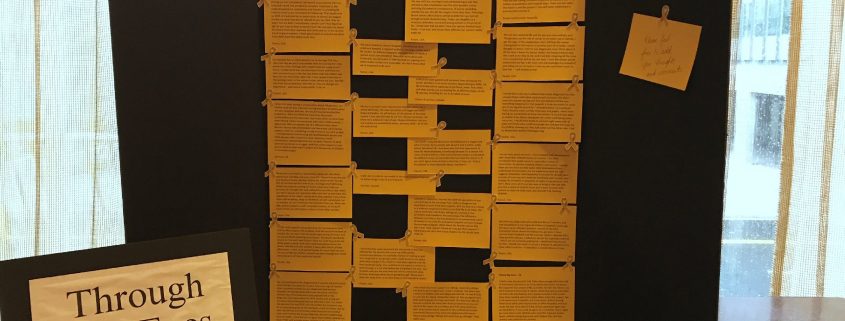
Through Our Eyes at One Retinoblastoma World 2017

Parents and survivors shared their thoughts on the “Through Our Eyes” wall at the One Rb World meeting in Washington D.C., 9-11 October 2017. These powerful insights were gathered anonymously via this website during September 2017, and highlight wide-ranging concerns.

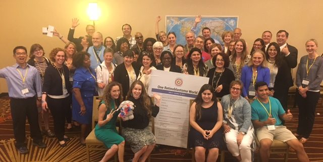
A large, diverse group of people pose together in a conference room. Most are dressed in smart casual attire. Many are smiling and waving at the camera, and those in the centre are holding a large banner that reads "One Retinoblastoma World". A large world map features on the wall behind the group. At the front, two ladies sit close together holding a plush elephant with removable eye between them. The elephant is bedecked in red, white and blue flowers.

One Retinoblastoma World 2017, Washington D.C.

Focused on best care for all, One Rb World 2017 included the first ever US national Family & Survivor Day. The collaborative program covered awareness, treatment, parent advocacy, life-long care, research, child life and the pathway to progress for our global community.

Stronger Together: Patient Engagement in Retinoblastoma Research

The Canadian Retinoblastoma Research Advisory Board is working with patient families to guide future research priorities. Engaging parents and survivors can lead to improved health outcomes. Read on and sign up today.

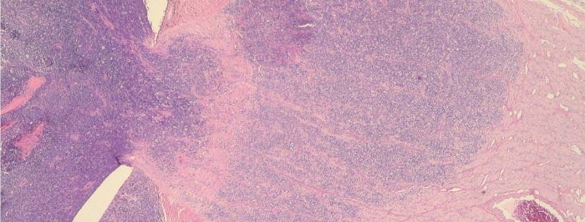
Pathology slide showing advanced retinoblastoma

The Anatomy of Retinoblastoma Pathology

Expert retinoblastoma pathology identifies cancer spread to outer layers of the eye or the optic nerve, guiding post-operative care.  WE C Hope CEO, Abby White explains how accurate, timely pathology is key to long-term survival when a child has advanced cancer, and what may happen when pathology is unreliable, or not available at all.

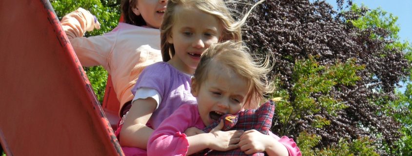
Three young girls sit one behind the other as they enjoy riding together down a slide. The girl at the back has curly brown hair and wears a light pink sweater. The middle girl has blonde hair and wears a light purple shirt. The girl in front has light brown hair, wears a pink top, and holds a plaid cushion. They are surrounded by green foliage and trees in the background. The sun creates halos around their hair as it blows with their movement down the slide, and blue sky is seen in the top right corner, beyond the trees..

Parents and Survivors Can Help Doctors Create Great Retinoblastoma Care

Being the parent of a child with retinoblastoma, or living with the effects of this cancer, can be frustrating. We look at a few challenges and ways we can help the medical community advance care.

A fuzzy cloud shows recurrence of a previously treated tumour.

Visualizing Cancer Treatment

Have you ever wondered what happens in a child’s eye during retinoblastoma treatment? We explore four of the most common eye-saving treatments here. Below each, you will find a link to further information that includes advantages, disadvantages, side effects and questions to ask the doctor.

This image has two sections. The top portion is a pictorial description of the TNM Staging, and comparison with the IIRC (comparison in brackets). Six images show progression of cancer through the different TNM stages, from small tumours confined to the retina in T1a (A) and T1b (B) to large tumours and seeds in T2a (C/C) and T2b (C/D). These first four images are grouped as “intraocular”. T3a-e (E) shows distress to tissue surrounding the eye. This image is indicated as high risk for extraocular spread. T4 (no IIRC comparison) shows an eye that has ruptured completely. This last image is described as extraocular. The lower section of the image shows average age at diagnosis in developed and developing countries, and the timeline to extraocular spread. The smiling child on the left is pictured with white pupil. The caption below reads: “North America: Unilateral Dx mean 27 mo | Bilateral Dx mean 15 mo”. The child on the right has a completely ruptured eye. The caption below reads “Kenya: Unilateral Dx mean 36 mo | Bilateral Dx mean 26 mo”. An arrow points from the left to right photo and reads “6 to 11 mo DELAY dx”.

Staging Intraocular Retinoblastoma

Retinoblastoma is staged to help doctors describe the degree of cancer in a child’s body, define potential for cure / eye salvage, determine best treatments, and compare impact of different therapies. Each eye is staged separately to define potential for safely saving the eye and vision.

Poster showing a child with one eye glowing white and the other, red. Caption above reads "a white glow in a child's eye could be a sign of cancer". Instructions below describe the PhotoRED technique.

How Do We Achieve Early Diagnosis of Retinoblastoma?

Retinoblastoma specialists agree that early diagnosis of eye cancer is a child’s best hope of survival, retaining some vision or their eye. Yet around the world, thousands of children die, and more than half of children who are cured lose at least one eye. We consider the potential and limitations of six solutions to improve early diagnosis.

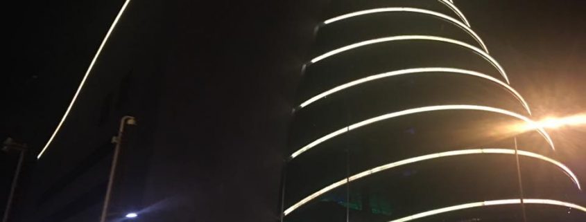
Dublin Convention Centre was lit up gold every night during the SIOP World Congress, celebrating the childhood cancer gold ribbon.

Alfred Knudson Memorial Session

A special session at the 2016 World Congress of Paediatric Oncology was hosted in memoriam of Alfred G. Knudson, Jr., MD, PhD, who died in July. Knudson developed his “two-hit hypothesis” after years of observing children with retinoblastoma, and this now forms the backbone of cancer genetics. The session focused on most effective staging for retinoblastoma, identifying and treating high risk children.

SIOP Welcome Signs

SIOP World Congress 2016

The International Society of Paediatric Oncology World Congress promotes a holistic approach to care. Through a unique collaboration of multiple diverse organizations, the program embraces almost every element of childhood cancer care and survivorship. Dublin hosted an inspired SIOP 2016 congress, October 19-22, a global village with a united voice and energy for all children and their families.