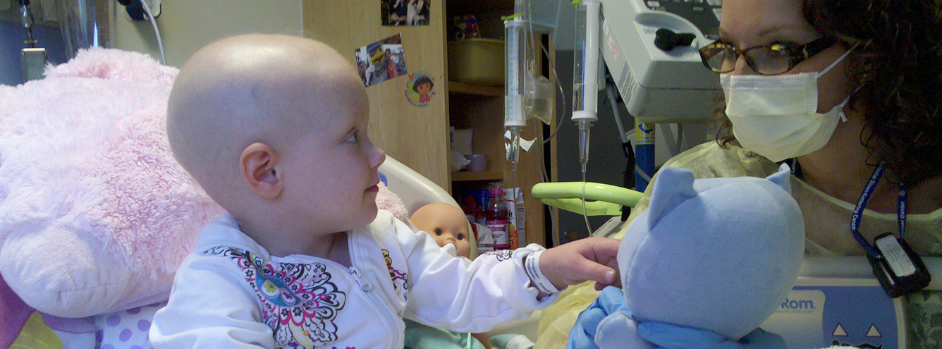
A chest x-ray makes pictures of the heart, lungs, airways, blood vessels and the bones of the spine and chest.
The images are displayed on a film or computer screen.
A chest X-ray is done:
- before chemotherapy begins
- to check placement of a central line
- if your child has a chest infection or poor breathing.

As x-rays involve radiation, they are not recommended for routine imaging in children or adults with a known or suspected constitutional RB1 mutation. Radiation exposure increases second cancer risk in these individuals.
Before the X-ray
The x-ray scanner is usually a wall-mounted, box-like machine containing the x-ray film or a special plate to record digital images. An x-ray producing tube is located about six feet away.
The patient may lie on a table over which the x-ray producing tube is suspended. The x-ray film or digital recording plate is housed in a drawer under the table.
Chest x-rays are painless and non-invasive, but your child may feel discomfort from the cool imaging plate. If she is very nervous about the x-ray, it may be possible to arrange a pre-test visit to familiarise her with the equipment.
Chest x-rays take about 20 minutes, including set-up and imaging. Your child will be asked to remove clothing or jewellery that can interfere with the imaging. She may also be asked to wear a hospital gown. The technician will then position her for each x-ray.
Two images will be taken. For the first view, your child will be positioned with hands on hips and chest pressed against the imaging plate. For the second, her side is against the image plate with arms elevated. Infants and young children will be positioned lying down on the table.
During the X-ray
The technician leaves the room during each x-ray. Many hospitals allow parents to stay, and you have the right to request this. Do advocate to stay with your child as your presence can be very reassuring. You will be asked to wear a lead apron to protect you from radiation exposure.
The x-ray machine emits a small burst of radiation that passes through your child’s body, recording an image.
Different parts of the body absorb x-rays to varying degrees.
- Bone and dense tissue absorbs more radiation, appearing lighter on the image.
- Soft tissue allows more radiation to pass through, and appears in shades of grey.
- Air appears black.
Your child may be asked to hold her breath for a few seconds while the x-ray is taken, to avoid blurred images.
After the X-ray
You will be asked to wait in the department until the technician has reviewed the images to ensure they are sufficient.
Chest x-ray images are available almost immediately. A radiologist will review them and send a signed report to your child’s doctor.