Ultrasound of the eye and orbit aids diagnosis of eye cancer and monitoring of treatment response.
An ultrasound is a quick and safe way to examine the back of your child’s eyes, where retinoblastoma forms.
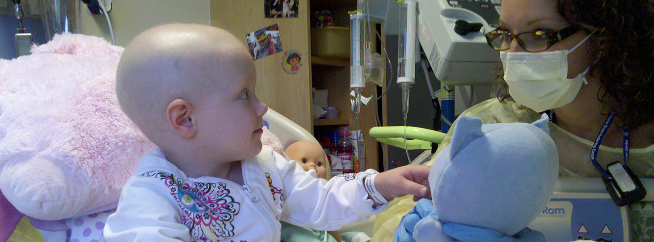
Ultrasounds are frequently done during EUA. When the child is awake, they are done either in the ophthalmology clinic or ultrasound (echography) department.

The test takes about 20 minutes, possibly longer if your child needs more time and support to complete the procedure.
There are two types of ultrasound scan for the eye, A and B. The B scan is used for children with retinoblastoma. The A scan may be used before cataract surgery following radiotherapy.
Anaesthetic Drops
Anaesthetic drops will be used to numb the eye. The drops feel uncomfortable and may sting, and this can upset children. The discomfort passes in about 30 seconds.
Preparing your child and giving lots of encouragement is very helpful. For specific suggestions on how to help your child when giving eye drops, click here to visit our Child Life section.
Try to discourage your child from rubbing her eyes for 20 minutes after the scan – she can accidentally scratch the cornea while the eye is still numb.
During the Procedure
Your child will sit in a chair a little like a dentist’s chair, or held in your arms. The doctor or ultrasonographer (a person trained in using ultrasound) will squirt a thick, clear lubricating gel on the surface of the eye, and place a handheld probe (transducer) on top of the gel.
You can gently remove gel residue with a warm wet cloth once the scan is complete.
The probe emits high frequency sound waves that travel through the eye and bounce off structures within it. The echoes of the sound waves reflect back to the probe, to form an image of the inner eye. For this reason, children often call the probe a “picture wand”.
The probe is moved around the surface of the eye, and your child will be asked to look in different directions to image the whole eye. Your child will feel some pressure from the probe. This sensation can be upsetting if the child is not prepared for it.
The images appear on a computer screen where video and still frames can be captured for detailed analysis later.
After the Procedure
The technician will clean the gel from your child’s eye and surrounding skin. You can gently remove gel residue with a warm wet cloth.
You will usually be asked to wait in the department until the ultrsonographer or doctor has reviewed the images to make sure they are sufficient.
Preparing for the Gel
The lubricating gel does not hurt but some children do not like the sensation of it, especially if very sensitive to being touched in the eye area. Help your child make a coping plan to ease her anxiety over this part of the procedure.
Daisy and her child life specialist, Morgan, wrote a silly song to prepare her for the ultrasound gel which she hated. Sung to the tune of Jingle Bells, the words are:
Slip and slide
Slip and slide
On the special jelly
Cold and wet is how it feels,
to help them see inside!